


Interventional Cardiology
95009 97678
rangsknathan@yahoo.com
1st Prize in Quiz Programme on Echocardiogram, conducted during Echo Workshop. Madras Medical Mission, Chennai.
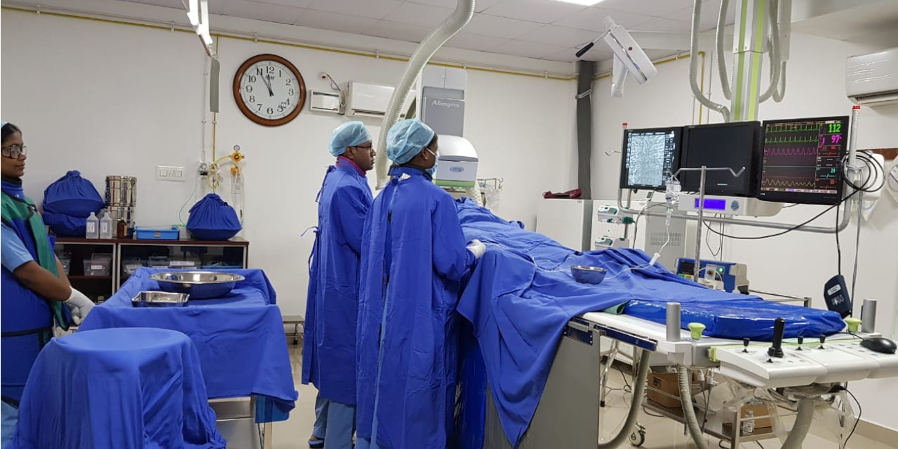
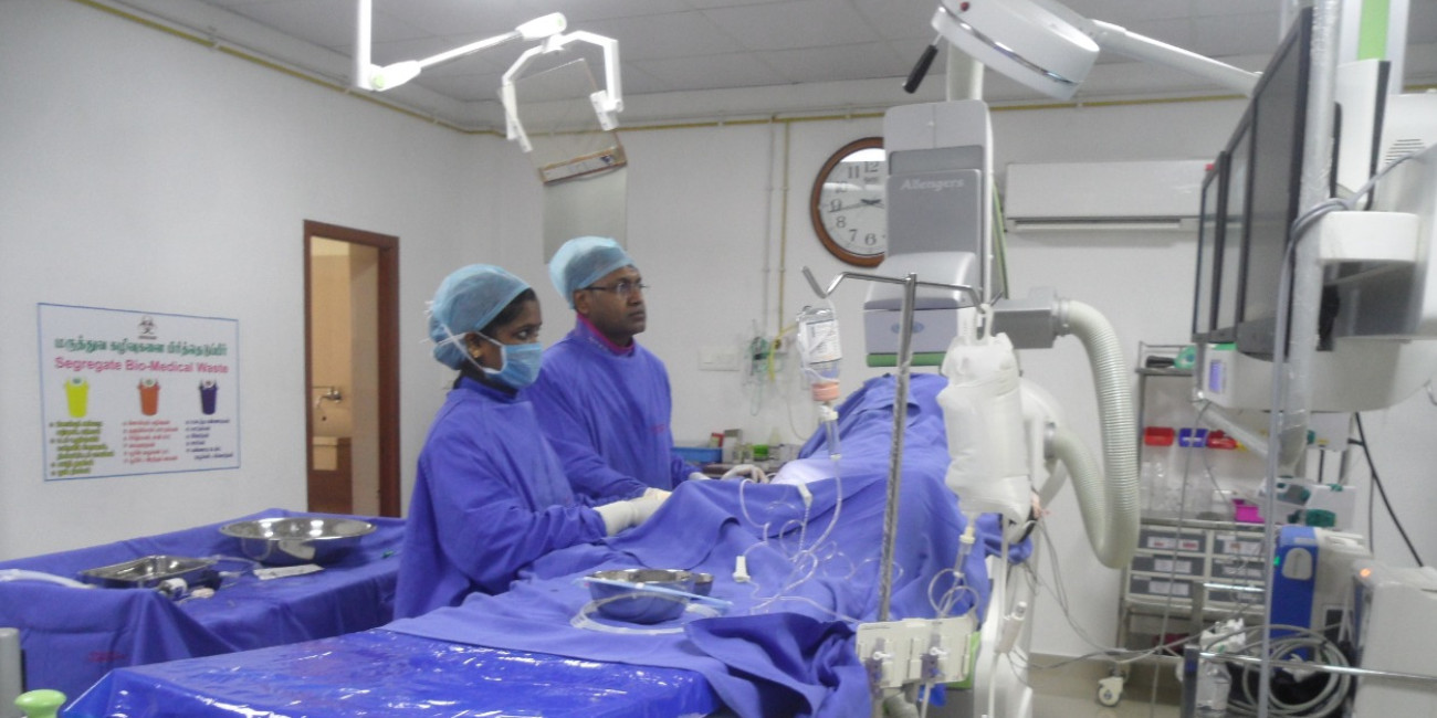
Dr. K. Ranganathan is a well-qualified Interventional Cardiologist with extensive training in his field of specialisation in India and the United Kingdom. He is currently practising in Pranav Hospitals, Brindhavan Road, Salem and AVM Hospital, Seelanayakkan patti, Salem.
Between obtaining his MD (General Medicine) postgraduate degree in 2001 and the DM (Cardiology) super speciality degree (2009-2012), Dr. K. Ranganathan trained in the UK from June 2002 to August 2007. He gained valuable experience there in the areas of General Medicine, Cardiology and other Medical specialities, serving in the capacity of Medical Registrar.
Subsequent to obtaining the DM degree, Dr. K. Ranganathan worked as Registrar, Interventional Cardiology, at Meenakshi Mission Hospital in Madurai, before joining as Consultant Interventional Cardiologist at Manipal Hospital in February 2013. He has considerable experience with interventional cardiac procedures. He has done over two thousand coronary angiograms (both radial and femoral approach), and about 500 angioplasties.
Dr. K. Ranganathan has attended numerous workshops and short courses to enhance his medical knowledge and skills. He is a regular participant at Cardiology conferences in the country, where he has presented his papers and research studies, some of which have been published in peer-reviewed journals.
Overview

The aorta is the major blood vessel that supplies blood to the body. An abdominal aortic aneurysm (AAA) is an enlarged and weakened area in the lower part of the wall of the aorta. A normal aorta is approximately one inch or less in diameter and it runs from the heart passing through the center of the chest and abdomen.
In general, aneurysms can develop anywhere along the length of aorta. Aneurysms in the upper part of the aorta are called thoracic aortic aneurysms. But, aneurysms are more common in lower parts of aorta and called as abdominal aortic aneurysms. These aneurysms may also be referred to as AAA or triple A.
An aneurysm can grow to be more than five inches in diameter. Due to high pressure of blood flowing through the artery, the weakened area enlarges like a balloon. It is more common to see large aneurysms bursting as compared to the smaller ones.
This results in internal bleeding that can lead to death unless treated immediately by an experienced cardiovascular surgeon. AAAs are considered a serious health condition because they can burst or rupture. Only about half of patients with a ruptured AAA who get to a hospital survive. Treatment of AAA may vary from watchful waiting to emergency surgery depending on the size and rate at it is growing. Once a diagnosis of AAA is made, doctors will closely monitor it so that surgery can be planned if it's necessary. It is very risky to wait for surgery till the time AAA ruptures.
Risk factors for abdominal aortic aneurysm include:
Causes
Aneurysms of aorta are more common in the abdominal part of aorta. The exact cause of abdominal aortic aneurysms is unknown, but various factors may play a role, which include:
Symptoms
Abdominal aortic aneurysms are mostly slow growing and have no symptoms. This makes them difficult to detect. Some aneurysms will never rupture. Many aneurysms are small to begin with and stay small, although many expand over time. Some expand quickly. It is very difficult to predict how fast an abdominal aortic aneurysm may enlarge.
Enlargement of an abdominal aortic aneurysm may lead to:
Any person who is a smoker or aged 60 years or more, with a family history of abdominal aortic aneurysm is at risk of developing an abdominal aortic aneurysm. He/she should consider regular screening for the condition. As male sex as well as smoking has preponderance for abdominal aortic aneurysm, men ages 65 to 75 who have ever smoked cigarettes should have a one-time screening for abdominal aortic aneurysm using abdominal ultrasound.
Diagnosis
Diagnosis of abdominal aortic is often by chance in patients presenting for examination for another disease. A pulsating bulge in abdomen may be felt by doctor during a routine exam. Aortic aneurysms are often found during routine medical tests. An X-ray of the chest or ultrasound of the heart or abdomen, sometimes ordered for a different reason, may lead to the diagnosis.
In case of high suspicion of an aortic aneurysm, the doctor may order specialized tests to confirm it. These tests might include:
Several medical bodies active in preventive medicine recommend that men aged 65 to 75 who have ever smoked should have a one-time screening for abdominal aortic aneurysm using abdominal ultrasound. The need for a screening ultrasound should be discussed with doctors by people older than age 60 with a family history of abdominal aortic aneurysm or other risk factors.
Complications
The main complication of abdominal aortic aneurysm is tears in the wall of the aorta (dissection). Life-threatening internal bleeding can ensue of an AAA ruptures. The risk of rupture is greater in large aneurysms.
Signs and symptoms indicating that aortic aneurysm has burst are:
Development of blood clots is another complication of aortic aneurysms. Small blood clots can develop in the area of the aortic aneurysm. A loose clot that breaks away from the wall of an aneurysm can block a blood vessel elsewhere in the body, causing pain or blocking the blood flow to the legs, toes, kidneys or abdominal organs.
Treatment
Treatment of AAA is very specific. Some general guidelines for treating abdominal aortic aneurysms are:
In case of a patient having a small abdominal aortic aneurysm — about 1.6 inches, or 4 centimeters (cm), in diameter or smaller — and without symptoms, the doctor may suggest a watch-and-wait (observation) approach, rather than surgery. Surgery, in general, isn't needed for small aneurysms because the risk of surgery likely outweighs the risk of rupture. If a patient chooses the observation approach, the doctor will monitor the aneurysm with periodic ultrasounds, usually every six to 12 months and encourage the patient to report immediately if there is abdominal tenderness or back pain — potential signs of a dissection.
The size of a medium aneurysm is between 1.6 and 2.1 inches (4 and 5.3 cm). How the risks of surgery versus waiting stack up in the case of a medium-size abdominal aortic aneurysm, is unclear. The benefits and risks of waiting versus surgery will need to be discussed with the doctor and then an informed decision be made with the help of the doctor. In case of watchful waiting, an ultrasound will be needed every six to 12 months to monitor the aneurysm size.
Surgery is generally required in cases of an aneurysm that is large (larger than 2.2 inches, or 5.6 cm) or growing rapidly (grows more than 0.5 cm in six months). Additionally, a leaking, tender or painful aneurysm requires treatment.
For abdominal aortic aneurysms, two types of surgeries are available:
Lifestyle measures are the best approach to prevent an aortic aneurysm as they keep the blood vessels as healthy as possible. That means taking these steps:
In case somebody has any of the risk factors for aortic aneurysm, it is very important to talk to the doctor. If you are at risk, your doctor may recommend additional measures. These include medications to lower blood pressure and relieve stress on weakened arteries.
Overview

Reduced blood flow to heart muscles causes chest pain called as angina pectoris. Angina is a symptom of coronary artery disease. A person may feel pain when insufficient oxygen-rich blood reaches the heart muscle. This reduced blood flow is caused by coronary artery disease. In coronary artery disease, there is an accumulation of plaque inside the coronary blood vessels.
The pain associated with angina is typically described as squeezing, pressure, heaviness, tightness or pain in your chest. A common presentation as told by most patients is the feeling that 'someone is standing on their chest'. The usual site of pain in angina is chest, but may also be felt in the shoulders, arms, neck, throat, jaw, or back.
Anginapectoriscan be a recurring problem or a sudden, acute health issue. Although angina is common, it can be hard to distinguish from other types of chest pain. Most common confounding factor is pain or discomfort of indigestion. Seek medical attention immediately if you have unexplained chest pain.
Causes
Reduced blood flow to your heart muscles causes angina. Blood is a carrier of oxygen, which your heart muscle needs to survive. In situations where heart muscle isn't getting enough oxygen, it causes a condition called ischemia. Coronary artery disease (CAD) is the most common cause of reduced blood flow to your heart muscles. The arteries of the heart (coronary arteries) can become narrowed by fatty deposits called plaques. This is called atherosclerosis.
Angina due to reduced blood flow is a supply problem, wherein the heart is not getting enough oxygen-rich blood. It might be interesting to note that a person may not always have angina if the heart arteries are narrowed due to fatty buildup. The reason is that during times of low oxygen demand — when you're resting, for example — your heart muscle may be able to get by on the reduced amount of blood flow without triggering angina symptoms. When the demand for oxygen suddenly increases, like when you exercise, this can cause angina.
A positive family history of CAD or stroke indicates increased risk to develop unstable angina than in people whose families do not have these conditions. Other risks for unstable angina are related to lifestyle, including:
Symptoms
Symptoms of angina include:
The typical features of chest pain and discomfort common with angina are a felling of pressure, squeezing, fullness or pain in the center of your chest. The feeling experienced by some people is of as if someone is squeezing their chest, or feeling like a heavy weight has been placed on their chest.
There are variations in the severity, duration and type of angina. In case somebody has a new or changing chest pain, immediate medical attention should be sought. New or different symptoms may signal a more dangerous form of angina (unstable angina) or a heart attack.
Most common form of angina is stable angina. It typically occurs with exertion and goes away with rest. When somebody previously healthy experiences a new onset chest pain, it's important to see the doctor to find out what's causing the chest pain and to get proper treatment. If your stable angina gets worse or changes, seek medical attention immediately. Characteristics of stable angina
Characteristics of unstable angina (a medical emergency)
Characteristics of variant angina (Prinzmetal's angina)
Symptoms of angina in women can be different from the classic angina symptoms. For example, a woman may have chest pain that feels like a stabbing, pulsating or sharp form of chest pain rather than the more typical vise-like pressure. Associated symptoms like nausea, shortness of breath or abdominal pain are more common in women. These differences may lead to delays in seeking treatment.
Diagnosis
to diagnose angina, your doctor will start by doing a physical examination and asking about your symptoms. You'll also be asked about any risk factors, including whether you have a family history of heart disease.
There are several tests your doctor may order to help confirm whether you have angina:
Complications
Heart attack is the most dangerous complication to be concerned about with angina. Common symptoms of a heart attack include:
Seek emergency medical consultation if you have any of these symptoms.
Treatment
Angina treatment options are many, including lifestyle changes, medications, angioplasty and stenting, or coronary bypass surgery. Treatment aims to reduce the frequency and severity of your symptoms and to lower your risk of heart attack and death.
Medications
Medications are required if lifestyle changes alone don't help your angina. Medications may include:
Overview

Blood vessels that carry oxygen and nutrients from the heart to the rest of the body are called arteries. Arteries that are not diseased are flexible and elastic. With increasing age and due to other factors, too much pressure in your arteries can make the walls thick and stiff — sometimes restricting blood flow to your organs and tissues. Hardening of arteries ensues which is called as arteriosclerosis.
Atherosclerosis is a specific type of arteriosclerosis, but the terms are sometimes used interchangeably. Buildup of fats and cholesterol in and on walls of arteries (plaques) is called as atherosclerosis. This process can restrict blood flow.
Another hazard associated with these plaques is that they can burst, triggering a blood clot. Atherosclerosis is generally considered a problem of arteries of heart, though it can affect arteries anywhere in your body. It is important to recognize that atherosclerosis is a preventable and treatable condition.
With increasing age, hardening of the arteries occurs. Apart from getting older as a risk factor, other factors that increase the risk of atherosclerosis include:
Causes
Atherosclerosis starts in the arteries as early as childhood and is a slow, progressive disease. The precise causes that lead to atherosclerosis are unknown. But, damage or injury to the inner layer of an artery may initiate the process of atherosclerosis. The damage may be caused by:
Damage to the inner arterial walls leads to build up of blood cells and other substances at the injury site and further damaging the inner lining of the artery. With passage of time, fatty deposits (plaques) made of cholesterol and other cellular waste products also build up at the injury site. The hardening and narrowing of your arteries is caused by these plaques. Blocked arteries lead to insufficient blood supply to the organs and tissues connected to themleading improper function of these arteries.
Further, these plaques may break off and enter bloodstream. Additionally, the smooth lining of a plaque may rupture, spilling cholesterol and other substances into your bloodstream. As a result of these changes a blood clot forms which can completely block the blood flow to a specific part of your body. If this happens in arteries of heart, it leads to a heart attack. It is also noteworthy that a blood clot can travel to arteries of other parts of your body and block (partially or totally) blood flow to another organ.
Symptoms
The process of atherosclerosis is a gradual one. Mild atherosclerosis usually doesn't have any symptoms. The symptoms of atherosclerosis don't develop until an artery is so narrowed or clogged that it can't supply adequate blood to your organs and tissues. In extreme circumstances, a blood clot blocks blood flow completely, or even breaks apart and can trigger a heart attack or stroke.
Moderate to severe atherosclerosis can lead to symptoms dependingupon which arteries are affected. For example:
If somebody feels he/she has above symptoms pointing towards atherosclerosis, it is important to meet a doctor. One should also be vigilant about early symptoms of inadequate blood flow, such as chest pain (angina), leg pain or numbness. The patients must understand that early diagnosis and treatment can stop atherosclerosis from worsening and prevent a heart attack, stroke or another medical emergency.
Diagnosis
During a physical examination, the doctor may find signs of narrowed, enlarged or hardened arteries. These include:
Complication
Location of atherosclerosis determines the complications of atherosclerosis. For example:
Treatment
The best treatment for atherosclerosis is lifestyle changes that include eating a healthy diet and exercising. But sometimes, medication or surgical procedures may be recommended as well.
Many drugs are available these days that can slow — or sometimes even reverse — the effects of atherosclerosis. Some commonly used drugs are:
More aggressive treatment is required sometimes.Surgical procedures are employed if you have severe symptoms or a blockage that threatens muscle or skin tissue survival. These include:
Lifestyle changes can help you prevent or slow the progression of atherosclerosis.
Overview

The human heart beats at a particular rate and with a particular rhythm. When there is a problem in either the rate or rhythm of the heart beat or when both are disturbed, the condition is called arrhythmia. So an arrhythmia could be due to heart beating too fast (tachycardia) or too slow (bradycardia) or with an irregular rhythm (for example extra or premature beats).
While some arrhythmias could be harmless, arrhythmias could turn serious if they affect the pumping action of the heart. to understand arrhythmia better, you need to understand how the heart works.
Electrical signals originate in a group of specialized cells called pacemaker node, located in the upper right chamber of the heart (right atrium). In a resting healthy heart, pacemaker node generates 60-100 electrical impulses in a minute. These impulses are conducted via a special conducting system to the right and left atria (upper chambers) which contract to pump blood into the right and left ventricles, respectively (lower heart chambers).
The electrical signal then reaches across and down the length of the heart and ventricles through specialized conducting pathways to ultimately activate the left and right ventricles. The right and left ventricles contract to push out blood to lungs and rest of the body, respectively. After this contraction, the ventricles relax, and a fresh impulse is generated at the pacemaker node.
Causes
Arrhythmias occur if there is a block or delay in the electrical pathways, irregular signal generation from pacemaker node, irregular impulse conduction or due to impulse generation in any part of heart except the pacemaker node. This could happen:
The four main types of arrhythmia are extra or premature beats, supraventricular arrhythmias, ventricular arrhythmias, and bradyarrhythmias.
These are the most common types of arrhythmia. They are of two types depending on where the extra betas occur: premature atrial contractions (PAC) and premature ventricular contractions (PVC).
Both PAC and PVC are usually harmless, can occur without a cause or due to stress, exercise, caffeine or nicotine intake, and need no treatment. There are no signs and symptoms except a feeling of flutter in chest or extra heart beat. At times they occur in heart disease and need treatment.
Supraventricular arrhythmias are basically tachycardias or tachyarrythmias originating in the atria or AV node. Examples: atrial fibrillation, atrial flutter, and paroxysmal supraventricular tachycardia (PSVT), congenital (Wolff-Parkinson-White syndrome)
In atrial fibrillation electrical impulse is generated anywhere in the atria or pulmonary veins and travels through out the atria in a disorganized manner. The atria quiver or fibrillate in a fast and irregular way instead of contracting properly.
Cause: Usually occurs due to coronary heart disease, rheumatic heart disease or high blood pressure, in hyperactive thyroid gland, with advanced age, or in alcohol abuse. Sometimes no cause is identified.
Complications: When rate of quivering becomes as 300 per minute or more, it may cause quivering of ventricles and heart failure. Blood pools in quivering atria and forms clots, which can break and travel to brain causing stroke.
This is similar to atrial fibrillation, except that the electrical impulses travel in an organized manner.
PSVT occurs when the signals go from atria to ventricles and come back to atria causing extra beats and short spurts of very fast heart rates. PSVT can occur during physical activity or without a cause in young people. Usually it is not dangerous.
In another PSVT type in Wolff-Parkinson-White syndrome, electrical impulses travel from the atria into the ventricles along an extra pathway as well.
Ventricular arrhythmia start in the ventricles, are usually serious and require urgent medical care. They occur in heart attack, coronary heart disease, and conditions causing weak heart muscles. There are two main types of ventricular arrhythmias:
Ventricular tachycardia is spurts of very fast but regular beating of ventricles. It is the less serious type of ventricular arrhythmia, and can become dangerous only if it lasts long.
In V-fib, highly disorganized electrical signals make the ventricles quiver or fibrillate instead of contracting properly. This can be extremely dangerous as it can result in sudden cardiac arrest and death. This needs urgent treatment by electrical shock therapy through a defibrillator.
In addition to above mentioned causes, it can occur if you suffer from potassium, calcium, and magnesium imbalance due to kidney disease or as effect of certain medications.
Bradyarrhythmias are heart rates slower than 60 beats per minute. This can be a normal finding in active adults. However, bradyarrhythmia can occur due to electrolyte imbalance, heart attack, underactive thyroid or use of drugs such as beta blocksrs, digoxin, calcium channel blockers.
Symptoms
Arrhythmias can occur without signs and symptoms or cause mild ones such as:
Serious symptoms of arrhythmia include:
Diagnosis
It is often difficult to diagnose arrhythmias as their symptoms occur once in a while and may be difficult to detect. Your doctor can diagnose arrhythmia by taking your medical and family history, doing a physical examination and certain tests.
Your doctor would:
During your physical examination, your doctor may:
Main Tests
Your doctor may or may not opt for these tests depending on your symptoms.
Treatment
Arrhythmias which cause symptoms and are serious can be treated with medication, medical devices and procedures, surgery and Vagal maneuvers. Serious arrhythmias are those which are likely to cause complications like stroke, heart failure and sudden cardiac arrest.
Medications used to treat arrhythmias are called antiarrhythmics. Some of these medications have serious side effects. You must take the medications as prescribed. Ask your doctor about their potential side effects. You must also immediately report any new symptom or recurrence of an old symptom.
Antiarrhythmic medications can:
Apart from antiarrhythmics, your doctor may prescribe blood thinners or medicines to control thyroid disorders.
Simple exercises can slow down or stop abnormal heart rhythms by affecting the Vagus nerve. However, please do not do these exercises without consulting your doctor.
Vagal maneuvers which can help are:
Overview

Any disease of the heart muscle that interferes with the heart's ability to pump blood with sufficient force is called cardiomyopathy. Cardiomyopathy develops slowly and may produce no symptoms until the later stages, except when caused by viral infections. It is an uncommon disorder, accounting for only 1 percent of heart disease fatalities.
But, it is one of the more common causes of serious heart disease in younger people. Coronary artery disease causes cardiomyopathy in the elderly. Treatment may include medications, implantable devices or, in severe cases, a heart transplant depending upon the type of cardiomyopathy.
There are three types of cardiomyopathies including:
Causes
Cause of the cardiomyopathy is unknown in most cases. However, doctors are able to identify some contributing factors in some people. Possible causes of cardiomyopathy include:
Symptoms
There is a lot of variations in the symptoms of cardiomyopathy.
Possible symptoms of dilated cardiomyopathy:
Possible symptoms of hypertrophic cardiomyopathy are generally same as the symptoms of dilated cardiomyopathy. Rarely the first symptom may be fainting or even sudden death. It can also cause heart attack, usually during exercise.
Possible symptoms of restrictive cardiomyopathy are fluid accumulates in the legs and abdomen. This condition also can cause shortness of breath, especially during exertion.
Irrespective of type of cardiomyopathy you have, signs and symptoms tend to get worse unless treated. This worsening happens quickly in certain people, while in others, cardiomyopathy may not worsen for a long time.
Diagnosis
On a doctor visit, a physical examination of the patient will be conducted. A personal and family medical history will be taken, and patient askedabout symptom occurence — for example, whether exercise brings on your symptoms. You may need to undergo several tests if your doctor thinks you have cardiomyopathy, to confirm the diagnosis. These tests may include:
Many other blood tests, including those to check your kidney function and look for anemia and thyroid problems, might be done. Your doctor may order for checking your iron level. Increased iron in blood may indicate an iron overload disorder called hemochromatosis. Accumulating too much iron in your heart muscle can weaken it and cause cardiomyopathy.
Complications
Other conditions arising due to cardiomyopathy are:
Treatment
The management of cardiomyopathy aims to reduce your signs and symptoms, prevent your condition from worsening, and decrease your risk of complications. Treatment varies by which of the major types of cardiomyopathy you have.
In this condition, your doctor may recommend medications, surgically implanted devices or a combination of both. The medications you may be prescribed include:
In some cases, a special pacemaker is implanted that coordinates the contractions between the left and right ventricles (biventricular pacing). Drug therapy or an implantable cardioverter-defibrillator (ICD) may be options in people who may be at risk of serious arrhythmias.
In this condition, your doctor may recommend beta blockers to relax your heart, slow its pumping action and stabilize its rhythm. These medications also calcium channel blockers, such as verapamil. In hypertrophic cardiomyopathy medications are often the preferred treatment. You may need surgery or a medical device to treat your condition if medications don't work. Options include:
Septalmyectomy.It is a type of an open-heart operation in which the surgeon removes part of the thickened, overgrown heart muscle wall (septum) that separates the two bottom heart chambers (ventricles). This removal improves blood flow and reduces mitral regurgitation. Most patients improve with this surgery and have no further symptoms.
Septal ablation.In this treatment, a small portion of the thickened heart muscle is destroyed by injecting alcohol through a catheter into the artery supplying blood to it. There are possible complications with this procedure, including heart block — a disruption of the heart's electrical system — which requires implantation of a pacemaker. Although long-term success of this procedure isn't yet known, it's becoming more commonly used.
Pacemaker implantation.In this procedure, a small electronic device is inserted under your skin that sends electrical signals to your heart to monitor and regulate your heartbeat. This procedure is generally done under local anesthesia and typically takes less than three hours. But, this procedure is not as effective as surgical options, but it's sometimes used in older people who want to avoid more invasive procedures.
Implantable cardioverter-defibrillator (ICD).In this procedure, a pager-sized device is implanted in your chest like a pacemaker. This device continuously monitors your heartbeat. In case a life-threatening arrhythmia is detected, the ICD delivers precisely calibrated electrical shocks to restore a normal heart rhythm. In patients at risk of a sudden cardiac death because of abnormal heart rhythms, this device is very useful.
The management of this condition aims at improving symptoms. The advice you will receive from doctor will recommend you to pay careful attention to your salt and water intake and monitor your weight daily. Diuretics might be recommended if sodium and water retention becomes a problem. Blood pressure lowering medications also ,may be prescribed to lower your blood pressure and control fast or irregular heart rhythms.
A heart transplant may be an option if you have severe cardiomyopathy and medications can't control your symptoms. Even people who are critically ill may have a long wait before having a heart transplant because of the shortage of donor hearts. A mechanical heart assist device can help In some patients who are critically ill as they wait for an appropriately matched donor. These devices, known as ventricular assist devices (VADs), can help blood circulate through your heart for months or even years.VAD therapy could be a long-term treatment option in some people who aren't candidates for a heart transplant.
Choose the following lifestyle measures to help you manage cardiomyopathy:
Specific recommendations will depend on the type of cardiomyopathy you have.
Generally, you can't prevent cardiomyopathy in many cases. If you have a family history of the condition, consult a doctor. If cardiomyopathy is diagnosed early, treatments may prevent the disease from worsening. Preventing the diseases that cause cardiomyopathy is the best to preventit. Know your risk factors for coronary artery disease and modify those risks early in life. You can reduce your risk for coronary artery disease by:
Contact your doctor for evaluation if you have any family members with inherited cardiomyopathy.
Overview

Coronary arteries are the major blood vessels that supply your heart with blood, oxygen and nutrients. Coronary artery disease (CAD) occurs when these arteries become damaged or diseased. The cause mainly is cholesterol-containing deposits (plaque) on your arteries. Narrowing of the arteries occur when plaques build up, causing your heart to receive less blood. The decreased blood flow, eventually, may cause chest pain (angina), shortness of breath, or other coronary artery disease signs and symptoms. Heart attack occurs when there is a complete blockage.
CAD can go virtually unnoticed until you have a heart attack because coronary artery disease often develops over decades. It can lead to angina or acute myocardial infarction if left untreated. However, one can actually do a lot to prevent and treat coronary artery disease, starting from committing to a healthy lifestyle. Treatment for coronary artery disease can include medications, or surgical and minimally invasive procedures.
There are many risk factors for CAD which includes:
In some cases, CAD develops without any classic risk factors. Researchers are studying other possible factors, including:
Causes
Damage or injury to the inner layer of a coronary artery initiates CAD, sometimes as early as childhood. The damage may be caused by various factors, including:
Damage to the inner wall of an artery leads to fatty deposits (plaques) made of cholesterol and other cellular waste products to accumulate at the site of injury in a process called atherosclerosis. Rupture of the surface of these plaques causes blood cells called platelets to clump at the site to try to repair the artery. Heart attack can occur due to this dumping.
Symptoms
Narrowed coronary arteries are unable to supply enough oxygen-rich blood to your heart — especially when it's beating hard, such as during exercise. Initially, the decreased blood flow may not cause any coronary artery disease symptoms. But, you may develop coronary artery disease symptoms as the plaques continue to build up in your coronary arteries. The symptoms are:
Diagnosis
Upon presentation to doctor, a medical history will be taken, a physical exam will be conducted routine blood tests will be ordered. Doctor may suggest one or more diagnostic tests as well, including:
Complication
Complications of coronary artery disease are:
Some of the complications of hypertensive retinopathy include:
Treatment
Treatment of coronary artery disease varies depending on the situation. The patient might be treated with medications, undergo an invasive procedure or both — depending on the severity of the condition and the amount of damage to heart.
More heart tissue loses oxygen and deteriorates or dies with each passing minute after a heart attack. Restoration of blood flow quickly is the main way to prevent heart damage. Medications given to treat a heart attack include:
Effects of PCOS can be offset by paying attention to the foods you eat and your activity levels:
Additionally, to medications, one of the following procedures might be done to treat your heart attack:
After these medications/procedure, the blood flow to your heart is restored and your condition is stable.
Lifestyle changes can help you prevent or delay the occurrence of heart attack due to coronary artery disease:
Overview

Hear Failure is a serious condition in which your heart fails to pump blood sufficient enough to meet your body's oxygen needs. It is also called as congestive cardiac failure, right side heart failure, left side heart failure or cor pulmonale. Heart Failure occurs when the heart cannot relax properly to fill in enough blood (right sided heart failure) or cannot contract properly to pump out enough oxygen rich blood (left sided heart failure) or due to a combination of both problems (congestive heart failure).
Right sided heart failure is a result of high blood pressure in pulmonary arteries (arteries taking blood from heart to lung) is called cor pulmonale.
As heart becomes weaker, toxins build up in the body, which worsen the symptoms.
Currently, there is no cure for heart failure. However, it can be managed by medications and life style changes to allow you to lead a normal and active life.
Heart failure is treated by cardiologists. However your regular doctor can help in diagnosis, and in managing your medication and treatment.
Heart has four chambers, two upper (right and left atrium) and two lower (right and left ventricle).
In normal condition the right atrium receives oxygen poor blood from the body, pushes it to right ventricle. From the right ventricle the blood is pushed to lungs through pulmonary arteries for purification. Oxygen rich blood from the lungs fills the left atrium. This blood is pushed to the left ventricle which contracts to push the oxygen rich blood to the rest of the body.
In left side heart failure, the heart fails to contract or relax properly. When the heart does not contract properly, less oxygen rich blood is pushed out to the body. The excess blood accumulates in the left ventricle and atrium. This creates a backward pressure and pushes the blood back into the lung. Thus fluid accumulates in the lung.
In right side heart failure, the right atrium and ventricle fail to relax properly. Hence they are unable to take the oxygen poor blood from rest of the body. This causes a backward pressure such that fluid accumulates in the body, especially in feet, ankle, legs and abdomen.
Usually both right and left sided heart failures are present at the same time. However one may be more prominent than the other.
Causes
The three common causes of heart failure are
Other Causes:
Conditions which damage heart muscle:
Symptoms
The signs and symptoms common in all types of heart failure are:
Diagnosis
Early diagnosis and treatment improves the life span and quality of life.
The diagnosis is made on the basis of medical and family history, physical examination and certain tests.
Your doctor would like to know if you or any family member has a history of heart disease, diabetes or high blood pressure. You will be asked to explain your symptoms in detail, when they started, how they progressed etc. Do your best to answer the questions to the best of your ability.
During the physical examination, your doctor will:
There is no single test specific for heart failure. After taking your medical and family history and physical exam, your doctor may ask for one or more of the following tests:
Treatment
The treatment of heart failure is aimed at keeping the symptoms in check and preventing worsening of the condition. The treatment includes medication, lifestyle changes, and other treatments.
Your doctor will prescribe medications depending on your symptoms and results of your tests. Please take the medications as prescribed. Do not miss doses and do not self-medicate. If you feel the medication is not suiting you, please inform your doctor immediately.
The various medications prescribed for heart failure are:
Simple lifestyle changes along with medications will help you dramatically improve your symptoms and lead a better quality of life.
A Heart Healthy Diet: You can ask the dietician in your heath care team to make customized diet plans for you. Otherwise you can follow these simple tips.
Since you have excess fluid in your body and you may be on water pills, it is important to drink only as much fluid as allowed. Drinking too much or too less may worsen your symptoms. Avoid alcohol completely.
Overview

Hypertension, or high blood pressure, is the most common cardiovascular disorder affecting populations across the world. Blood pressure actually indicates the force of blood pushing against artery walls as it flows through the arteries or blood vessels in the human body. Blood vessels get filled by blood up to a certain capacity.
too much pressure of blood on the vessel wall, or high blood pressure, can threaten healthy arteries and lead to life-threatening conditions such as heart disease and stroke. High blood pressure is hazardous due to its propensity to cause strokes, heart attacks, heart failure, or kidney disease. Everybody should be aware about his/her blood pressure.
Generally, high blood pressure is categorized as:
Treatment strategies to manage high blood pressure include changing lifestyle suitably and possibly drug therapy to lower blood pressure to less than 140/90. For patients who have diabetes or chronic kidney disease the recommended blood pressure is less than 130/80. Aim of management is to lower high blood pressure and protect important organs, like the brain, heart, and kidneys from damage. Research has found that treatment for hypertension causes a significant reduction in stroke (reduced an average of 35%-40%), heart attack (20%-25%), and heart failure (more than 50%).
Causes
It is not known exactly what causes high blood pressure. But, there exist several factors and conditions that may play a role in its development. These factors include:
As per the guidelines the normal blood pressure should remain within 120/80 mm Hg. A very stringent life style modification is recommended in terms of diet control & modest exercise if the blood pressure ranges in between 120-139/ 80-89 mm Hg & medical treatment is recommended if the blood pressure is more than 140/90 mm Hg. High blood pressure tends to run in families and is more likely to affect men than women. Increasing age and race also play a role in causation. Diet and lifestyle also greatly affect es hypertension. Other factors that can raise the risk of having hypertension include obesity; diabetes; stress; insufficient intake of potassium, calcium, and magnesium; lack of physical activity; and chronic alcohol consumption.
Symptoms
Most of the patients may not even know that they have hypertension. The only way to know if your blood pressure is high is through regular checkups. Some patients do have certain symptoms, especially if their blood pressure is extremely high. These are:
In case anybody has these symptoms, a doctor should be consulted immediately. Hypertension that is not treated or poorly treated can lead to stroke, heart disease, kidney failure and eye problems.
Diagnosis
Hypertension is often called a "silent disease" as the patient usually doesn't know about it. There may be no symptoms or signs. But, it keeps on damaging your body. So, it's important to regularly monitor your blood pressure. Tests should be conducted for heart disease as hypertension is a risk factor for heart disease.
Blood pressure is measured in mm hg. Most often, blood pressure is measured with a device known as a sphygmomanometer. Blood pressure is measured in two ways: systolic and diastolic. Systolic blood pressure is the maximum pressure during a heartbeat. Diastolic blood pressure is the lowest pressure between heartbeats.
According to the most recent guidelines, a normal blood pressure is less than 120/80 mm Hg. Hypertension is blood pressure that is greater than 140/90, while prehypertension consists of blood pressure that is 120 to 139/80 to 89.
Apart from BP measurement, your doctor will ask about your medical history (whether you've had heart problems before), assess your risk factors (whether you smoke, have high cholesterol, diabetes, etc.), and talk about your family history (whether any members of your family have had high blood pressure or heart disease).
A physical examination of the whole body will also be conducted by your doctor. If you're diagnosed with high blood pressure, your doctor may recommend other tests, such as:
Complications
High blood pressure which is not managed or is uncontrolled can lead to:
Treatment
Any patient with BP readings greater than 120/80 should be encouraged to enroll for treatment planning. This includes lifestyle modifications, such as eating a healthier diet, quitting smoking, and getting more exercise. Treatment with medication is recommended to lower blood pressure to less than 140/90. For patients who have diabetes or chronic kidney disease the recommended blood pressure is less than 130/80.
Treating high blood pressure involves lifestyle changes and possibly drug therapy.
Medication
Your doctor may prescribe one or more prescription medicines to help lower your blood pressure in addition to taking steps such as losing weight and exercising. Most of the medications prescribed for treatment of hypertension help lower your chances of developing health problems such as stroke, kidney disease, or heart attack.
In these times, a lot of drugs from various categories of antihypertensives are available that can effectively help most people lower their blood pressure and reach the blood pressure goal set by their health care provider.
Different medications prescribed for control of blood pressure work in many different ways. Some remove extra fluid and salt from the body. Others slow down your heartbeat or relax and widen your blood vessels.
In market, there are more than 100 blood pressure–lowering drugs available. Some of them, like diuretics, have been in use for more than 50 years.
Many pills prescribed for high blood pressure contain a combination of two different types of medicines. Combining two different types of drugs can make it easier to control blood pressure by two different mechanisms. These combination pills also keep track of taking your medicines.
Do keep in mind that since combination pills contain more than one medicine, they also have the potential for more side effects than pills that contain just one medicine. Report anything unusual to your doctor while you are taking pills for hypertension.
Different types of medications work differently for different types of patients, with varying age and body profile. In fact, some medicines have been found to provide more benefits for men than women or younger than in older populations. Your doctor will provide a medicine that is right for you.
Reduction of blood pressure with medication will prevent progression from less severe to more severe hypertension, will prevent enlargement of the heart and heart failure, and will dramatically reduce strokes, stroke deaths, and heart attack deaths. Getting blood pressure down to below 140/90 mm Hg, or below 130/80 mm Hg if you have diabetes or kidney disease, if at all possible, is a good idea at all ages.
Do remember that even if you are taking your medications regularly, it is important to keep on exercising and eating as advised by your health care provider.
Overview

Heart attack, technically known as myocardial infarction (MI) or acute myocardial infarction (AMI), occurs due to diminution of blood supply to a part of the heart, causing that particular heart cells to die. Heart attack most commonly occurs due to occlusion (blockage) of a coronary artery following the rupture of a vulnerable atherosclerotic plaque.
It results into restriction in blood supply and oxygen shortage ensues. If this situation is left as such without treatment for a sufficient period of time, it can cause damage or death (infarction) of heart muscle tissue (myocardium).
Unwanted buildup of fatty deposits (atherosclerosis) that narrows arteries throughout your body, including arteries to your heart, is contributed by certain factors. It is possible to improve or eliminate many of these risk factors as this decreases your chances of having a first or subsequent heart attack.
Risk factors for heart attack include:
Causes
When one or more of the arteries supplying your heart with oxygen-rich blood (coronary arteries) become blocked, it results in heart attack. With passage of time, a coronary artery can become narrowed from the buildup of cholesterol called plaque. This process of plaque of buildup in arteries throughout the body is called atherosclerosis. One of these plaques can rupture during heart attack and a blood clot forms on the site of the rupture. A complete blockage of flow through artery occurs if the clot is large enough. Coronary artery disease occurs when your coronary arteries have narrowed due to atherosclerosis. Most heart attacks occur due to coronary artery disease.
Spasm of a coronary artery that shuts down blood flow to part of the heart muscle is another uncommon cause of a heart attack. Life-threatening spasm can occur due to drugs such as cocaine. A tear in the heart artery (coronary artery dissection) can also cause heart attack. Small blood clots or tumors that have traveled from other parts of the body (coronary embolism) are other uncommon causes of heart attack.
The heart attack is culmination of a several hours long process. More heart tissue is deprived of blood and deteriorates or dies with each passing minute. Damage to the heart can be limited or prevented if blood flow can be restored in time.
Symptoms
Most common symptoms of heart attack are:
Women patients have different or atypical signs and symptoms of heart attack, which include:
Symptoms of heart attack may vary. All patients of heart attack do not experience the same symptoms or experience them to the same degree. Another interesting feature is that many heart attacks aren't as dramatic as the ones you've seen on TV. In some patients, there are no symptoms at all. Still, the more signs and symptoms you have, the greater the likelihood that you may be having a heart attack.
The timing of heart attack is not fixed and it can occur at any time of the day— at work or play, while you're resting, or while you're in motion. In some patients heart attacks strike suddenly, but many people who experience a heart attack have warning signs and symptoms hours, days or weeks in advance. Most common and earliest warning of a heart attack may be recurrent chest pain (angina) that's triggered by exertion and relieved by rest. A temporary decrease in blood flow to the heart causes angina.
Diagnosis
If a patient is having a heart attack or is suspecting having one, his/her diagnosis will likely happen in an emergency setting. You will be undergoing certain tests that will help check if your signs and symptoms, such as chest pain, signal a heart attack or another condition. These tests include:
After taking immediate steps to treat your condition, doctor may order for these additional tests:
Complications
Complications of heart attack are often related to the damage done to your heart during a heart attack. Most common problems arising due to damage to heart are:
Treatment
Treatment of a heart attack varies depending on the situation. The patient might be treated with medications, undergo an invasive procedure or both — depending on the severity of the condition and the amount of damage to heart.
Medication
More heart tissue loses oxygen and deteriorates or dies with each passing minute after a heart attack. Restoration of blood flow quickly is the main way to prevent heart damage. Medications given to treat a heart attack include:
Additionally, to medications, one of the following procedures might be done to treat your heart attack:
After these medications/procedure, the blood flow to your heart is restored and your condition is stable following your heart attack. You may be kept in the hospital for observation.
Lifestyle changes can help you prevent or delay the occurrence of heart attack.
Overview

When leg arteries become narrowed or blocked by plaque it causes peripheral Arterial Disease (PAD), a condition that raises the risk of heart attack, stroke, leg amputation and death. As patients are unaware of this condition generally, PAD remains under-diagnosed and under-treated.
In PAD, characteristically, there is a reduction in blood flow to the lower extremities due to plaque build-up in the leg arteries (also known as atherosclerosis). A combination of fats, cholesterol and other substances constitutes plaque. Size of plaque can grow to significantly reduce blood flow through an artery .
Blood flow to the legs and feet is reduced when leg arteries are hardened and clogged. Peripheral arteries remain blocked, completely or partially, and cause pain, changes in skin color and temperature, sores or ulcers and difficulty walking. PAD, if left untreated, can lead to Critical Limb Ischemia (CLI), a condition where not enough blood is being delivered to the leg to keep the tissue alive. Gangrene ensues due to total loss of circulation to the legs and feet leading to amputation. Hardened arteries, in addition, found in people with PAD are a sign that they are likely to have hardened and narrowed arteries to the heart and the brain. That is why people with PAD are at high risk for having a heart attack or a stroke. They may become disabled and not be able to go to work, with poor quality of life.
PAD is a chronic disease with risk factors that include:
Smokers or people with diabetes have the greatest risk of developing peripheral artery disease due to reduced blood flow.
Causes
Atherosclerosis is the main cause of peripheral artery disease. Fatty deposits (plaques) build up in your artery walls and reduce blood flow in atherosclerosis. This disease can and usually does affect arteries throughout your body although the heart is usually the focus of discussion of atherosclerosis. Atherosclerosis in the arteries supplying blood to your limbs causes peripheral artery disease.
The cause of peripheral artery disease, less commonly, may be blood vessel inflammation, injury to your limbs, unusual anatomy of your ligaments or muscles, or radiation exposure.
Symptoms
Generally, most people with peripheral artery disease have mild or no symptoms. Some people have leg pain when walking (intermittent claudication). The symptoms of intermittent claudication include muscle pain or cramping in your legs or arms that's triggered by activity, such as walking, but disappears after a few minutes of rest. Pain is localised to the area supplied by the clogged or narrowed artery. Pain in the calf is the most common location.
There is a wide variation in the severity of intermittent claudication, ranging from mild discomfort to debilitating pain. Walk or doing other types of physical activity could be a hard task due to severe intermittent claudication.
Other symptoms of peripheral artery disease include:
Progression of peripheral artery disease can lead to pain that may even occur when you're at rest or when you're lying down (ischemic rest pain). Its intensity could be strong enough to disrupt sleep. Temporary relief could be obtained by hanging your legs over the edge of your bed or walking around your room.
Diagnosis
to diagnose peripheral artery disease, your doctor may rely on the following tests:
Complications
Plaque buildup in blood vessels leading to peripheral artery disease (atherosclerosis) leads to a risk of developing:
Treatment
There are two major goals of treatment for peripheral artery disease. Managing symptoms, such as leg pain, so that you can resume physical activities is the primary goal. to stop the progression of atherosclerosis throughout your body to reduce your risk of heart attack and stroke is the secondary goal.
These goals can be accomplished with lifestyle changes. Quitting the smoking habit is the single most important thing you can do to reduce your risk of complications. You need additional medical treatment if lifestyle changes are not enough. Your doctor may prescribe medicine to prevent blood clots, lower blood pressure and cholesterol, and control pain and other symptoms.
Medication
In extreme cases, angioplasty or surgery may be necessary to treat peripheral artery disease that's causing intermittent claudication:
Your doctor may prescribe a supervised exercise training program, in addition to medications or surgery, to increase the distance you can walk pain-free. Regular exercise improves symptoms of PAD by a number of methods, including helping your body use oxygen more efficiently.
The symptoms of peripheral artery disease can be managed effectively with lifestyle measures and the progression of the disease can be slowed, especially by quitting smoking. to stabilize or improve PAD:
Care of the feet is very important in addition to the above suggestions. Follow this advice to care for your feet:
Maintain a healthy lifestyle to prevent claudication. That means:
An alternative approach to pacemaker implantation: A case report of pacemaker implantation through femoral access in an elderly lady with subclavian vein stenosis and pyopericardium. Medical E Journal of The Tamilnadu Dr. MGR Medical University.
A case report of pericardial effusion caused by ankylostomiasis. Indian Journal of Medical Microbiology.
Bald aortic arch in Takayasu arteritis: A case report of aorto arteritis. Postgrauate Medical Journal.
Review article on diastolic dysfunction. Cardiology Update 2009. 16th Annual Conference of the Indian College of Cardiology, Lucknow.
A rare case of aortic right atrial tunnel. 62nd Annual Conference of CSI. Dec 2010.
Comparative study of coronary angiogram by radial and femoral approach. 62nd Annual Conference of CSI. Dec 2010.
Prospective study of hypocalcemia in heart failure. 63rd Annual Conference of CSI. Dec 2011.
Incidental venous anomalies during cardiac intervention - a case series. 63rd Annual Conference of CSI. Dec 2011.
Pathophysiology of diastolic heart failure. Annual Conference of Tamil Nadu CSI, Yercaud. Oct 2013.